
http://openi.nlm.nih.gov/detailedresult.php?img=2892034_pone.0011293.g001&req=4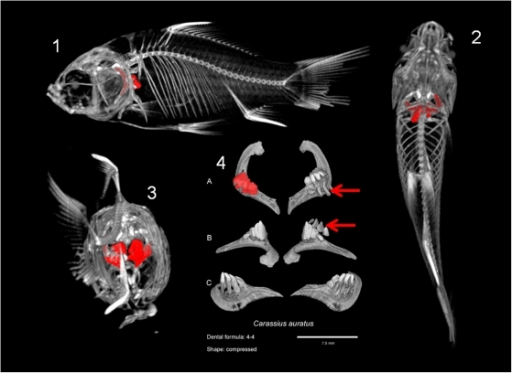 |
one-0011293-g001: Localization of the fifth ceratobranchial in the goldfish, Carassius auratus (Cyprinidae) and dental plate for this species.1, 2 and 3 are general views of the whole goldfish skeleton with the fifth ceratobranchial, bearing the pharyngeal teeth, painting in red. 1: Lateral view; 2: Ventral view; 3: Posterior view. 4 is the dental plate for this species with three conventional views: A: Occlusal view; B: ventral view; C: dorsal view. The dental formula for the goldfish is 4/4 (there is, on each side, one row of four teeth). Moreover, on one side, replacement teeth are visible as they are not attached to the pharyngeal bone (pointed by red arrows). The tooth shape is “compressed”.
Mentions: Figure 1 shows the example of a plate for a Cyprinidae species, Carassius auratus, with the orientation of the dentition within the animal body. All the plates for all the species are presented in Figure S1 and a selection of diverse morphologies is presented in Figure 2. From this dataset we extracted the dental formula and we qualitatively described the tooth shape with reference to various previously described morphotypes [4], [33]. In Cyprinoidea with several tooth rows, the tooth shape was determined for teeth on the ventral row as they are the biggest and the most differentiated. By rotating 3D reconstructions, it is easy to distinguish functional teeth from replacement teeth because the latter are not bound to the bone (see teeth pointed by red arrows on Figure 1).
|
Abstract: The fish order Cypriniformes is one of the most diverse ray-finned fish groups in the world with more than 3000 recognized species. Cypriniformes are characterized by a striking distribution of their dentition: namely the absence of oral teeth and presence of pharyngeal teeth on the last gill arch (fifth ceratobranchial). Despite this limited localisation, the diversity of tooth patterns in Cypriniformes is astonishing. Here we provide a further description of this diversity using X-ray microtomography and we map the resulting dental characters on a phylogenetic tree to explore evolutionary trends.We performed a pilot survey of dental formulae and individual tooth shapes in 34 adult species of Cypriniformes by X-ray microtomography (using either conventional X-ray machine, or synchrotron microtomography when necessary) or by dissecting. By mapping morphological results in a phylogenetic tree, it emerges that the two super-families Cobitoidea and Cyprinoidea have followed two distinct evolutionary pathways. Furthermore, our analysis supports the hypothesis of a three-row dentition as ancestral for Cyprinoidea and a general trend in tooth row reduction in most derived lineages. Yet, this general scheme must be considered with caution as several events of tooth row gain and loss have occurred during evolutionary history of Cyprinoidea.Dentition diversity in Cypriniformes constitutes an excellent model to study the evolution of complex morphological structures. This morphological survey clearly advocates for extending the use of X-ray microtomography to study tooth morphology in Cypriniformes. Yet, our survey also underlines that improved knowledge of Cypriniformes life traits, such as feeding habits, is required as current knowledge is not sufficient to conclude on the link between diet and dental morphology.


